tissue
Lung Immune Ecosystem
Air-exposed barrier where epithelial alarmins, macrophage tone, and type 2 circuits regulate airway reactivity
Review layer
Last reviewed 2026-05-17
Systems teaching draft. Content is structured for education and graph expansion, with formal source tagging ready for the next review pass.
Appears in response traces
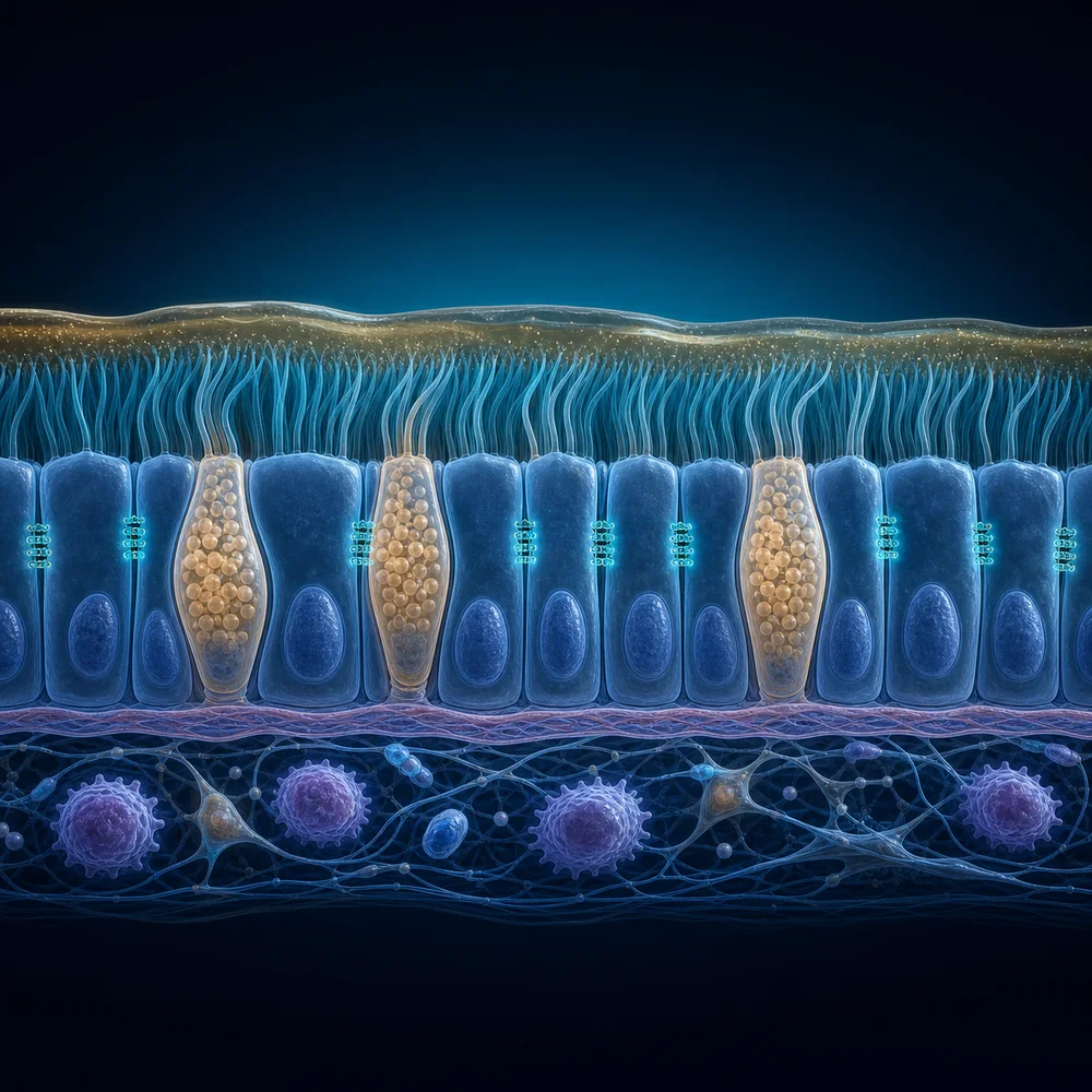 Educational illustration · not microscopy
Visual specimen
Airway epithelial barrier
The air-facing mucus and cilia layer sits above a joined epithelial sheet and a quieter immune tissue compartment.
Look for
- Mucus above coordinated cilia
- Goblet cells within the epithelial sheet
- Immune sentinels below the basement membrane
Conceptual tissue cutaway, not histology. Cell proportions, density, and spacing are simplified and not to scale.
OpenAI-generated educational illustration under Atlas editorial direction · 2026-07-12
State signature
Systems profile
System effects
Primary mapped axes from existing Atlas data.
Tissue
Organ, barrier, stromal, vascular, or local niche behavior.
Inflammation
Inflammatory alarm, recruitment, mediator release, or tissue-damaging amplification.
Tolerance
Immune restraint, resolution, regulatory tone, or set-point control.
Tissue ecosystem
Spatial immune niche
niche
Lung
Barrier
Cells
Cytokines
Neuroimmune
Graph neighborhood
Direct relationships
Arrow shows upstream source toward receiving target.
Airway-resident mast cells shape bronchial reactivity
Arrow shows upstream source toward receiving target.
Allergic airway type 2 loops
Arrow shows upstream source toward receiving target.
Airway epithelial injury and alarmin release
Arrow shows upstream source toward receiving target.
Mucus, airway reactivity, and tissue remodeling
Context link: no causal arrow.
Airway resident immune memory shapes recall, tolerance, and remodeling after inhaled exposures
Immune niche
Tissue Ecology
The lung balances inhaled antigen tolerance, antiviral readiness, allergic repair programs, and neutrophilic injury risk.