IMMUNE OS ATLASby AllerimSign in
← All journeys

02 · Tissue alarm to type 2 ecology

How does a stressed barrier become an allergic response loop?

Move from airway barrier sensing to IL-33, mast-cell activation, and the wider type 2 immune pattern.

10-14 minpublic educationno patient data
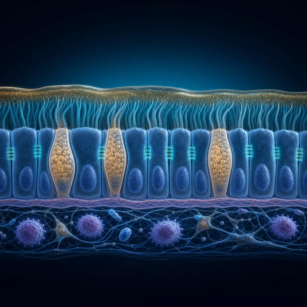
Educational cutaway of an intact airway barrier with mucus, cilia, goblet cells, epithelial junctions, basement membrane, and immune cells below.
Educational illustration · not microscopy

Visual specimen

Airway epithelial barrier

The air-facing mucus and cilia layer sits above a joined epithelial sheet and a quieter immune tissue compartment.

Look for

  • Mucus above coordinated cilia
  • Goblet cells within the epithelial sheet
  • Immune sentinels below the basement membrane

Conceptual tissue cutaway, not histology. Cell proportions, density, and spacing are simplified and not to scale.

OpenAI-generated educational illustration under Atlas editorial direction · 2026-07-12

Question to carry

How do tissue signals recruit and amplify an immune response instead of acting as isolated molecules?

Who this is for

A systems-immunology pathway for learners who want to connect tissue, signals, cells, and pattern.

Your journey progress

0 of 4 stops opened.

0% explored

Progress stays in this browser. Atlas records only deduplicated aggregate counters with no identifiers, free text, search terms, or patient data.

Follow the route

One connected story, 4 stops.

1

Begin in the tissue

Lung immune ecosystem

Start with barrier function, mucociliary clearance, resident cells, nerves, and environmental sensing in one spatial immune niche.

Boundary: The lung page describes a biologic ecosystem, not a person-specific airway state.

Open stop
2

Follow the alarm

IL-33 signaling

See how an epithelial or stromal alarm signal can lower activation thresholds and organize downstream type 2 behavior.

Boundary: A cytokine relationship shows plausible biology; it does not imply a measured cytokine level.

Open stop
3

Inspect the amplifier

Mast-cell relationship field

Trace how resident mast cells receive IgE, alarmin, complement, and neuroimmune context and produce rapid or sustained outputs.

Boundary: Multiple inputs can converge on similar symptoms, so mechanism requires context.

Open stop
4

Zoom out to the pattern

TH2 allergic ecology

Connect IL-4, IL-5, IL-13, IgE, eosinophils, mast cells, barrier tissues, and regulatory restraints as a dynamic pattern.

Boundary: A type 2 pattern is an educational lens, not a diagnosis or a treatment selector.

Open stop

What to take away

01

Barrier tissues actively sense and shape immune responses.

02

IL-33 is one signal inside a larger tissue and cellular network.

03

Type 2 immunity includes defense, repair, allergy, regulation, and tissue remodeling context.

Educational boundary

This journey is educational and teaches a general mechanism. It does not identify the cause of symptoms, distinguish emergencies, or recommend medication, testing, or treatment.

Evidence record

Versioned and source-anchored

Reviewed 2026-07-12 · Atlas editorial baseline

Owner Mark Pruitt · next review 2026-10-12 · stale on 2026-11-12

v1.0source anchoredreviewcontext requiredcurrent

Known uncertainty

Barrier signaling varies by tissue, trigger, timing, host state, and treatment context.

The pathway is deliberately simplified so the relationships remain legible.

Atlas Professional

Compare response states

Approved users can inspect the allergen response trace and organize competing pattern hypotheses in Atlas Professional.

Request professional access